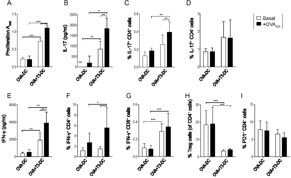
Fig. 5. T3-stimulated DCs induced a pro-inflammatory response in vivo. DCs treated with ovalbumin (100 μg/ml, OVA-DC) and 5 nM T3 (OVA+T3-DC) for 18 h, were injected i.v. into syngenic OTII transgenic mice. One week later, spleen cell suspensions were prepared and cultured with OVA323 peptide (50 ng/ml), and proliferation (A), IL-17A (B, C, D) and IFN-g production (E, F, G), CD4+CD25+FoxP3+ Treg cells (H) and PD-1-expressing CD4 cells (I) were determined 4 days later. Data are expressed as mean ± SD and are representative of 3 experiments with similar results performed in triplicate (4 mice per group). *p<0.05, **p<0.01, ***p<0.001.